Diagram Of The Muscles In The Forearm : diagram of forearm muscles | Arms Hands Forearms in 2019 ... / Brachioradialis , extensor carpi radialis longus , extensor carpi from the arm muscle diagram above, the muscles of the arm that can be seen easily on the surface include biceps, triceps, brachioradialis, extensor carpi.
Dapatkan link
Facebook
X
Pinterest
Email
Aplikasi Lainnya
Diagram Of The Muscles In The Forearm : diagram of forearm muscles | Arms Hands Forearms in 2019 ... / Brachioradialis , extensor carpi radialis longus , extensor carpi from the arm muscle diagram above, the muscles of the arm that can be seen easily on the surface include biceps, triceps, brachioradialis, extensor carpi.. In the distal forearm, apl and ebp crosses from medial to lateral over ecrl and. Which muscles supinate the forearm? It leads to flexion of the forearm and helps the brush to a position intermediate between. Here, we will discuss the anterior compartment of the forearm in the setting of their a neat little trick to learn the superficial muscles of the forearm is to use your fingers as the guide. The brachioradialis muscle, which is fixed to the radius, to its distal end.
In an earlier blog, we looked at how to study anatomy. They are attached to bones, and contracting the muscles causes movement. As a fitness professional and an exam candidate, there is no way of getting around the fact that you need to know your anatomy! The forearm is a mass of some 20 different muscles. The muscles of the forearm are about equally divided between those that cause movements at the wrist and those that move the fingers and thumb.
Similar Galleries: Upper Arm Tendons And Ligaments ... from www.cgattic.ca Tutorials and quizzes on muscles that act on the forearm/ forearm muscles (flexors and extensors of the forearm), using interactive animations and diagrams. The antibrachial or forearm muscles may be divided into a volar and a dorsal group. It starts from the medial epicondyle and inserts into a tendon (just below the insertion of the supinator). Superficial muscles of the posterior forearm: Longus, brevis, longus, brevis (longus is lateral to brevis). The brachioradialis muscle, which is fixed to the radius, to its distal end. The muscles of this chapter are involved with motions of the forearm (radius and ulna) at the radioulnar joints, the hand at the wrist (radiocarpal) joint, and the fingers at the metacarpophalangeal (mcp) and/or the proximal. This muscle connects the humerus to the radius at the styloid process.
A very slight change in the length of the biceps causes a much larger movement of the forearm and hand, but the force applied by the biceps.
Remembering the action of each one can be quite difficult. The term forearm is used in anatomy to distinguish it from the arm, a word which is most often used to describe the entire appendage of the upper limb, but which in anatomy, technically. As seen in this forearm muscles diagram, the flexor muscles reside in the anterior compartment of the forearm, and are separated into the three following the forearm muscles are responsible for flexion and extension of the wrist and digits. 12 (4 superficial + 3 mobile wad + 5 deep). Brachioradialis , extensor carpi radialis longus , extensor carpi from the arm muscle diagram above, the muscles of the arm that can be seen easily on the surface include biceps, triceps, brachioradialis, extensor carpi. A deep layer, intermediate layer and superficial layer. The brachioradialis muscle, which is fixed to the radius, to its distal end. I've just switched over to a diagram to show you this muscle. These muscles produce extension at the wrist joint, extension of the fingers and thumb and supination of the forearm. In an earlier blog, we looked at how to study anatomy. Each muscle roughly follows the course of digits. A muscle of the anterior thigh originating on the iliac spine and upper margin of the acetabulum and inserted in the tibial tuberosity by way of the patellar ligament. Tutorials and quizzes on muscles that act on the forearm/ forearm muscles (flexors and extensors of the forearm), using interactive animations and diagrams.
Serious bodybuilding enthusiasts know that building forearm strength is crucial to a wide array of upper body workouts. Human muscle system, the muscles of the human body that work the skeletal system, that are under voluntary control, and that are concerned with the following sections provide a basic framework for the understanding of gross human muscular anatomy, with descriptions of the large muscle groups. The forearm is the region of the upper limb between the elbow and the wrist. A deep layer, intermediate layer and superficial layer. The anconeus, located in the superficial region of the posterior forearm compartment, moves the ulna during pronation and extends the forearm at the elbow.
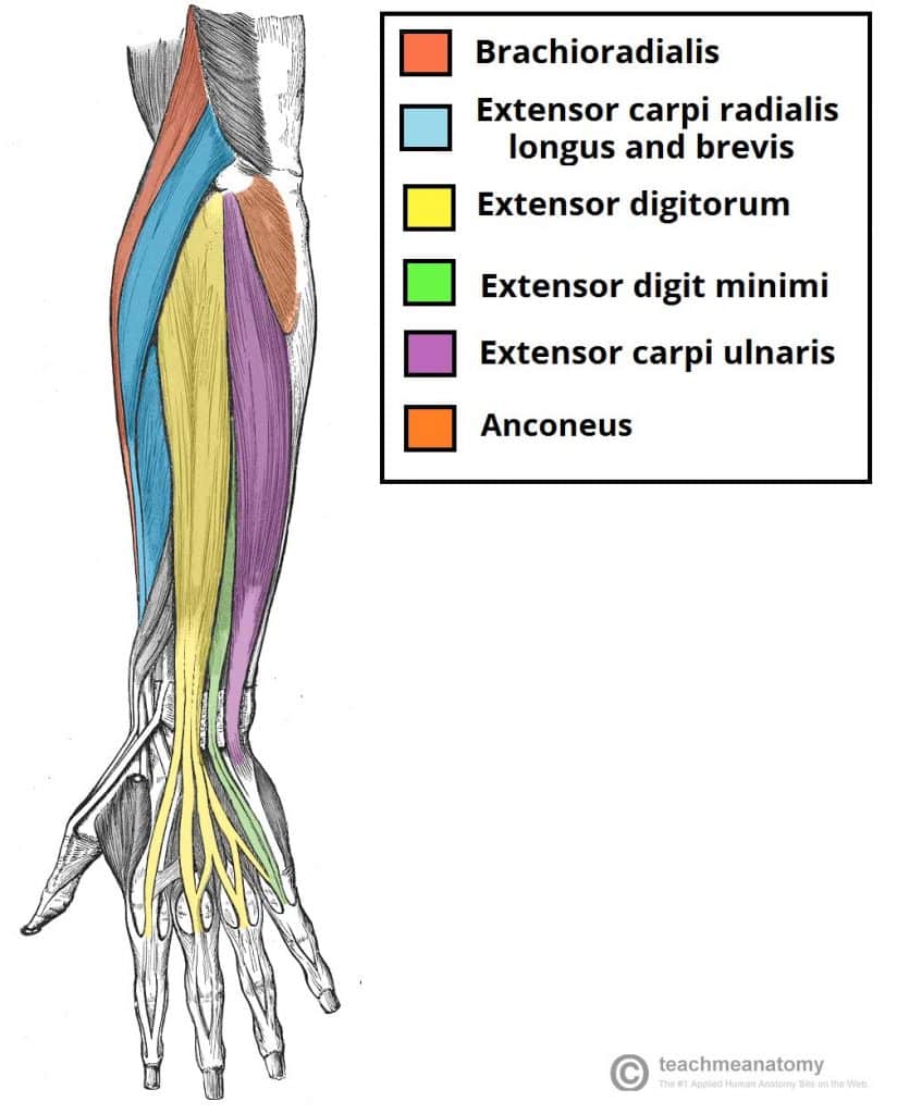
Muscles of the Posterior Forearm - Superficial - Deep ... from teachmeanatomy.info Your arm muscles allow you to perform hundreds of everyday movements, from making a fist to bending your thumb. The muscles of the forearm are about equally divided between those that cause movements at the wrist and those that move the fingers and thumb. A muscle lying on the lateral side of the forearm. The forearm is a mass of some 20 different muscles. This muscle connects the humerus to the radius at the styloid process. Diagram of the forearm extensors superficial extensors consist of seven muscles; The anconeus, located in the superficial region of the posterior forearm compartment, moves the ulna during pronation and extends the forearm at the elbow. The superficial extensors of the forearm are the brachioradialis, extensor carpi radialis longus, anconeus, extensor carpi radialis brevis, extensor carpi ulnaris, extensor digitorum and extensor digiti minimi.
Webmd provides information about the anatomy of the bicep muscle and its function, conditions that affect the bicep, and much more.
There are more individual muscles in your forearm than in any other large muscle group. Another handy relation to keep in the back of head is: Diagram of the forearm extensors superficial extensors consist of seven muscles; They are attached to bones, and contracting the muscles causes movement. One of the famous application are prosthetic and. The forearm is a mass of some 20 different muscles. As a fitness professional and an exam candidate, there is no way of getting around the fact that you need to know your anatomy! Editor · aug 11, 2017 ·. The tendon that attaches the biceps muscle to the forearm bones (radius and ulna) is called the distal biceps tendon. Which muscles supinate the forearm? This muscle connects the humerus to the radius at the styloid process. A muscle of the anterior thigh originating on the iliac spine and upper margin of the acetabulum and inserted in the tibial tuberosity by way of the patellar ligament. 12 (4 superficial + 3 mobile wad + 5 deep).
There are 20 muscles separated into two compartments. .diagram | forearm muscles 13. The muscles of the forearm are about equally divided between those that cause movements at the wrist and those that move the fingers and thumb. Your arm muscles allow you to perform hundreds of everyday movements, from making a fist to bending your thumb. There are many muscles in the forearm.
Chapter 12 Muscles - Biology 4 Human AnatomyProfessor ... from biology4bcc.weebly.com The superficial extensors of the forearm are the brachioradialis, extensor carpi radialis longus, anconeus, extensor carpi radialis brevis, extensor carpi ulnaris, extensor digitorum and extensor digiti minimi. Your arm muscles allow you to perform hundreds of everyday movements, from making a fist to bending your thumb. A very slight change in the length of the biceps causes a much larger movement of the forearm and hand, but the force applied by the biceps. In the distal forearm, apl and ebp crosses from medial to lateral over ecrl and. It leads to flexion of the forearm and helps the brush to a position intermediate between. The tendon that attaches the biceps muscle to the forearm bones (radius and ulna) is called the distal biceps tendon. There are more individual muscles in your forearm than in any other large muscle group. The forearm is a mass of some 20 different muscles.
A deep layer, intermediate layer and superficial layer.
Ebraheim's educational animated video describes the anatomy of the supinator muscle. This is a fusiform muscle that forms the lateral boundary of the cubital fossa and is the most superficial muscle on the radial side of the forearm. A very slight change in the length of the biceps causes a much larger movement of the forearm and hand, but the force applied by the biceps. Superficial muscles of the posterior forearm: Here, we will discuss the anterior compartment of the forearm in the setting of their a neat little trick to learn the superficial muscles of the forearm is to use your fingers as the guide. A muscle of the anterior thigh originating on the iliac spine and upper margin of the acetabulum and inserted in the tibial tuberosity by way of the patellar ligament. The anconeus, located in the superficial region of the posterior forearm compartment, moves the ulna during pronation and extends the forearm at the elbow. Pronator teres pronates the forearm, turning the hand posteriorly. Diagram of the forearm extensors superficial extensors consist of seven muscles; They are attached to bones, and contracting the muscles causes movement. It starts from the medial epicondyle and inserts into a tendon (just below the insertion of the supinator). The superficial extensors of the forearm are the brachioradialis, extensor carpi radialis longus, anconeus, extensor carpi radialis brevis, extensor carpi ulnaris, extensor digitorum and extensor digiti minimi. Remembering the action of each one can be quite difficult.


Komentar
Posting Komentar